An MRI of Robert Bowers’ brain shows an unusually large number of white matter lesions that could be indicative of schizophrenia, an expert testified Tuesday.
But those types of lesions also can be caused by uncontrolled high blood pressure, migraines or heavy smoking, the expert said.
Dr. Murray A. Solomon, a neuroradiologist based in California, was one of five physicians called by the defense Tuesday to support its argument that Bowers is ineligible for the death penalty because mental illness and brain abnormalities rendered him incapable of forming the requisite intent.
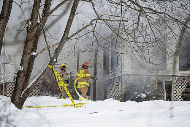
Earlier this month, a jury found Bowers guilty of all 63 federal counts against him for the Oct. 27, 2018, attack at the Tree of Life synagogue in Squirrel Hill that left 11 congregants dead. Tuesday was the second day in the eligibility phase of the trial.
To find that Bowers is eligible for the death penalty, the government must prove intent and at least one of four aggravating factors, including that the defendant created a grave risk of death to 12 additional people, that the crime involved substantial planning and premeditation, that those killed were considered “vulnerable victims” because of their age and intellectual disabilities, or that there were multiple killings.
The victims, shot and killed during Shabbat services, were members of the Tree of Life-Or L’Simcha, Dor Hadash and New Light congregations. They included Rose Mallinger, 97; Bernice Simon, 84, and her husband, Sylvan Simon, 86; brothers David Rosenthal, 54, and Cecil Rosenthal, 59; Dan Stein, 71; Irving Younger, 69; Dr. Jerry Rabinowitz, 66; Joyce Fienberg, 75; Melvin Wax, 87; and Richard Gottfried, 65.
During opening statements in the trial’s eligibility phase Monday, defense attorney Michael Burt told jurors that brain imaging completed on his client — including an MRI, PET scan and EEG — was consistent with schizophrenia and epilepsy.
On Tuesday, Solomon was the last witness to testify. He appeared via videoconferencing on monitors set up throughout the courtroom.
Working as a neuroradiologist since 1983, Solomon testified that defense attorneys asked him to review an MRI completed on Bowers by UPMC on Jan. 3, 2022.
At the time of the request, Solomon said he was not aware of any previous diagnoses and learned only a few weeks ago — as he was preparing to testify and read other expert reports — that schizophrenia and epilepsy were at issue in Bowers’ case.
Solomon testified that Bowers’ MRI showed a number of abnormalities, including asymmetry in the left and right hippocampus, which is where new memories are formed, and 28 white-matter lesions, which he called “highly unusual for a 49-year-old male.”
The lesions, Solomon said, are scars in the brain and the affected areas no longer function. He described images from the MRI showing the small spots and said that having a large number of the lesions can affect cognition, reasoning and decision-making.
“These lesions are everywhere throughout the brain, throughout the hemispheres,” Solomon said.
The asymmetry in the hippocampus, Solomon said, is worrisome and could be indicative of permanent brain damage.
“It could have been caused by trauma or a seizure disorder,” he said. “It’s a red flag.”
U.S. Attorney Eric Olshan noted during cross-examination that the report Solomon wrote in the Bowers case doesn’t include any reference to schizophrenia.
Solomon testified that he did not know schizophrenia could cause an increase in white-matter lesions until a few weeks ago.
“Then I Googled it because I was in disbelief,” Solomon said.
When he did, a number of results were returned.
“I researched it, I read articles,” he said.
Solomon said he also didn’t consider schizophrenia as a cause of the lesions when he wrote his report because he did not know Bowers had previously been diagnosed with it.
“Schizophrenia is a relatively unusual diagnosis,” Solomon said. “We don’t usually bring it up.”
The defense has not yet presented any evidence of Bowers’ underlying diagnosis, who made it or when.
Solomon’s testimony followed that of UPMC neuroradiologist Dr. Joseph Mettenburg, who conducted the initial review of Bowers’ brain MRI.
He also said he noticed extensive “white-matter hyperintensities,” which he noted could also have been caused by a number of things, including migraines or sickle cell disease.
He called Bowers’ brain imaging “a normal brain MRI,” and not diagnostically helpful.
While there were no abnormalities, Mettenburg said, “Plenty of MRIs for people with epilepsy are normal.”
Relative to Bowers’ PET scan, the defense called Dr. Andrew Newberg, a nuclear medicine physician at Thomas Jefferson University in Philadelphia.
Newberg testified that he is an expert in reviewing PET scans and has written more than 250 articles during his career.
In the Bowers case, the defense asked Newberg to review a PET scan completed in November 2021 to see if it would match another expert’s findings of schizophrenia.
A PET scan measures blood flow and glucose processing in the brain. Newberg, also testifying via videoconference, told the jury that he observed significant abnormal activity in the defendant’s scan, including an overactive frontal lobe and general asymmetry in the brain.
The overactive frontal lobe, Newberg said, can be caused by inflammation and can lead to a persistently excited state.
Newberg said an overactive, increased metabolism in the frontal regions of the brain is associated with problems concentrating, processing information, organizing and planning, emotional dysregulation, and impaired initiation and generation of thoughts.
Newberg testified that he also saw abnormally decreased metabolism in Bowers’ left medial temporal lobe, which can be associated with impaired memory, abstract reasoning and a variety of delusions, including religious delusions.
Bowers also had an abnormally decreased metabolism in his hypothalamus, which Newberg said regulates the autonomic nervous system and fight-or-flight response. It can lead to an inappropriate stress response, excessive reactions and misperceived threats.
Newberg said his findings were consistent with extensive clinical abnormality, including problems with emotional regulation, cognitive impairment, processing problems and psychiatric symptoms, including schizophrenia.
Newberg said that his comparisons were based on FDA-approved software that look at the results of PET scans from other people who are healthy.
On cross-examination by Olshan, Newberg said that the software’s control group database only includes 43 people, and just 20 in Bowers’ age range.
Newberg also admitted during cross-examination that a PET scan by itself cannot be used to diagnose anything and cannot be used to predict behavior — including whether a person can form the intent to kill.
The initial UPMC radiologist to read Bowers’ PET scan, Dr. James Michael Mountz, said he was not asked about schizophrenia, but instead was tasked with finding an “epileptogenic focus” — or imaging in the brain indicating symptoms of epilepsy.
He didn’t find one.
“Can you have a negative MRI and a negative PET scan and still have epilepsy?” Burt asked.
“Yes,” Mountz answered.
But, under cross-examination by Olshan, Mountz said that if a patient had no clinical history of seizures and clean MRIs and PET scans, it is possible they do not have epilepsy.
“You don’t scan patients to find epilepsy,” Mountz said. Instead, he said, you scan them to find the focus and manage their symptoms.
Also testifying Tuesday was UPMC neurologist and epilepsy specialist Dr. Vijayalakashmi Rajasekaran. She reviewed data from an “ambulatory,” or outpatient, EEG performed on Bowers on Dec. 14, 2021. An EEG measures the electrical activity of the brain.
Rajasekaran testified that Bowers’ EEG indicated he had a “potential seizure tendency,” but he did not have any seizures during the testing. Rajasekaran estimated that she has reviewed 5,000 to 6,000 EEGs in her nine years as an epilepsy specialist.
Brain waveforms seen in an EEG should be slow and soft, but Bowers’ brain waveforms in the imaging were “sharply contoured,” Rajasekaran testified. That is sometimes indicative of a patient with seizures, she said.
Rajasekaran said the test alone cannot tell whether the patient has had a seizure in the past or predict them in the future.
“It is very common not to capture a seizure” in an EEG, she said.
All three of the UPMC physicians said their reviews of Bowers’ brain imaging were inconclusive as to whether he has epilepsy. None of them were asked to interpret the results with respect to schizophrenia.
The defense will resume calling witnesses on Wednesday.